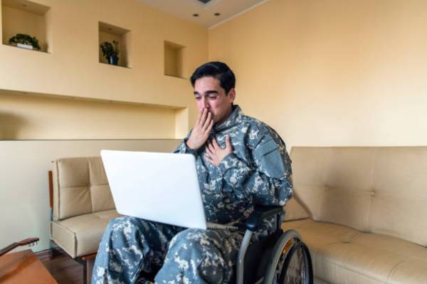
UAB Study Finds 30-Year Diagnostic Tool for Brain Injuries Unreliable

A landmark University of Alabama at Birmingham study, published in the American Journal of Roentgenology [radiology] in May 2025, and noted by UAB News on September 17, 2025, shows that “White matter hyperintensities [are] no longer a reliable way to diagnose mild traumatic brain injury”. Challenging a diagnostic method used for over three decades, the study found that white matter hyperintensities (WMH)—bright spots visible on brain MRI scans—appear equally in patients with mild traumatic brain injury and healthy individuals, undermining their diagnostic value.

Since the 1990s, WMH have been used to detect subtle brain damage invisible in standard imaging. The 1993 study A semiquantative rating scale for the assessment of signal hyperintensities on magnetic resonance imaging found on the NIH database, established the credibility of WMH by correlating these MRI findings with actual brain tissue damage, including demyelination and axonal degeneration. This finding has been largely accepted in the medical field, as shown by various studies since, many of which reported by the NIH, and made WMH attractive for diagnosing concussions when conventional scans appear normal.

However, medical professionals increasingly questioned the approach. New clinical reports show that WMH lack specificity—appearing not only from trauma, but also from normal aging, silent strokes, multiple sclerosis, and vascular disease. The UAB study confirmed these concerns: 34% of brain injury patients showed WMH compared to 35% of controls, with no correlation between WMH presence and symptom severity or recovery outcomes. Lead researcher Dr. Manoj Tanwar concludes that WMH alone is not enough for a diagnosis and comprehensive clinical assessment remains essential beyond imaging.

A Broader View of Diet’s Role in TBI Recovery

I tend to post about particular foods and diets that aid in brain injury recovery and overall brain health. This week, I take a broader view and examinine the benefits that one’s general choices of sustenance have in recovery and wellbeing:

In a holistic sense, research reveals that what survivors eat may dramatically influence their recovery. A groundbreaking 2023 NIH report published in Current Physical Medicine and Rehabilitation Reports found that specific dietary patterns can significantly improve long-term outcomes for brain injury patients. The study concluded that, “a diet rich in fiber and nutrients, but limited in added sugars, saturated fats, and excess calories would likely have the greatest cardiovascular and related neurologic protection.”

This finding was confirmed last year, with the Department of Defense and Veterans Affairs officially acknowledging in 2024 that, “nutrition may be a modifier of mild traumatic brain injury sequelae.” The research examined Mediterranean, DASH, and MIND diets, all emphasizing fruits, vegetables, healthy fats, and omega-3 fatty acids. These dietary interventions offer hope for reducing cardiovascular complications and improving neurological outcomes without pharmaceutical interventions.

Recently, in July 2025, the Journal of Neurotrauma published the findings of a study conducted by UNC School of Medicine, the Uniformed Services University, and the NIH that singled out a debilitating side effect of brain injury. They confirmed the importance of diet in preventing headaches, as trials demonstrated, “dietary changes significantly reduce persistent post-traumatic headaches…”

As can be seen, a comprehensive diet overhaul, with the possible assistance of a medical professional, can be a great benefit to both brain injury survivors and the general population.

The Connection Between TBI & Brain Cancer

On September 23, 2025, the University of Missouri School of Medicine reported groundbreaking findings that adults with moderate-to-severe traumatic brain injury face a 50% increased risk of developing brain cancer. The cancer is defined by malignant tumors that grow rapidly and invade surrounding brain tissue. Brain cancer affects approximately 24,820 Americans annually, according to the American Cancer Society, with a 33% five-year survival rate. Importantly, brain cancer itself is classified as an acquired brain injury—a form of non-traumatic brain damage that disrupts brain function.

The relationship between brain injury and brain cancer involves a fascinating interplay. While brain cancer causes acquired brain injury through tumor growth, research now suggests moderate-to-severe traumatic brain injuries may trigger brain cancer development through inflammation synergizing with existing genetic mutations. Not all brain tumors are cancerous, though. Approximately 72% are benign, even if they are still potentially serious depending on location.

Dr. Nimish Mohile of the University of Rochester notes that, “we’re starting to see progress with targeted therapies in the field of neuro-oncology,” offering hope for improved outcomes. Current NIH Director, Dr. Monica Bertagnolli, highlights that, “an AI-based diagnostic system can determine in just 10 seconds if part of a cancerous brain tumor that could be removed still remains,” demonstrating technological advances in treatment.

Prevention strategies include wearing helmets, preventing falls, and maintaining workplace safety—the same measures that protect against head injuries may reduce brain cancer risk. For those diagnosed with brain cancer, treatment often mirrors brain injury rehabilitation, as well—physical therapy, occupational therapy, speech therapy, and cognitive rehabilitation. These comprehensive approaches help patients regain function and improve quality of life, with research showing brain tumor patients recover at rates comparable to stroke and traumatic brain injury patients.

The encouraging news is that while moderate-to-severe TBI increases risk, the absolute probability remains low, and advances in early detection and treatment continue to improve outcomes. Still, patients with TBI history may benefit from monitoring for early tumor detection.

Brain Food in Peak Season

While beetroot peak harvest season is from late summer through October, research demonstrates that its vibrant juice offers remarkable benefits to the brain year round, specifically for brain injury and trauma recovery.

Studies published on the NIH databases and other reputable medical sources, such as that conducted by the University of Exeter and published by Free Radical Biology and Medicine journal in 2025, demonstrate how beetroot juice enhances cerebral blood flow and neuroprotection. “Our findings suggest that adding… beetroot juice – for just ten days can substantially alter the oral microbiome for the better, maintaining healthy brain function and slowing negative vascular changes,” explains Professor Anni Vanhatalo from the University of Exeter.

The scientific benefits of beetroot center on its high nitrate content—up to 11.4 grams per liter—which dramatically improves blood flow to injured brain tissue. Wake Forest University’s 2010 research first proved that beetroot juice increases blood flow to vulnerable brain regions, with researchers documenting enhanced oxygen delivery crucial for neural recovery.

Research at the University at Buffalo from 2023 also showed that beetroot juice can improve cerebrovascular function during stress, noting improved “cerebral autoregulation” mechanisms that protect against brain damage. Stroke recovery research shows 18 stroke survivors experienced increased nitrate levels supporting neural healing after 30 days of supplementation.

Digital Double-Edged Sword for the Brain

With over 4.9 billion active users globally spending an average of 2.5 hours daily on social media platforms, concerns about users’ neurological impact are mounting.

Parental and professional concern is warranted, it seems.  Social media platforms continue promoting risky challenges that can cause brain injuries. For example, the recent “Run It Straight” challenge on TikTok, YouTube, and Facebook involves participants sprinting toward each other and colliding at full force, creating what experts call “engineered systems for brain injury” with impacts similar to that of unbelted car crashes.

The worry, though, goes beyond risky behavior. The landmark July 8, 2025 NIH study “Modern Day High: The Neurocognitive Impact of Social Media Usage” found that social media use causes marked alterations in brainwave activity, with Beta and Gamma waves heightened during engagement and continuing after use, which potentially interferes with emotional regulation and attention.

Statistically, 74% of people with traumatic brain injury (TBI) use the internet. While this may be a lesser amount, compared to 84% of the general population, social media can be of greater risk for the brain injured population. Specifically, those with brain injuries face unique challenges navigating digital spaces due to cognitive impairments affecting memory, attention, and decision-making.

The growing concern has been felt in the government, too. Former Surgeon General Dr. Vivek Murthy recently warned, “Congress has not stepped up to its responsibility to protect our kids. They need to act now” regarding social media’s impact on developing brains. In the past few years, there has been an increase in funding for research into digital wellness and brain health initiatives targeting vulnerable populations. However, if, and to what level, the government should be involved in an individual’s social media activity is very much up for debate.

Yet social media isn’t entirely harmful. Rehabilitation professionals report that it can reduce social isolation for brain injury patients and support community reintegration. Still, “ongoing robust research is urgently required to give rehabilitation professionals an evidence-based framework” for safe implementation.

Your Brain and Lungs Talk to Each Other—And That Could Save Lives

Groundbreaking research published in September 2025 by the University of Miami Miller School of Medicine reveals that brain injuries send deadly molecular messengers throughout the body that directly attack lung tissue. The team discovered the “neural-respiratory inflammasome axis,” where injured brains release tiny particles called extracellular vesicles that travel through blood and trigger lung cell death.

Up to 30% of brain injury patients develop acute lung injury, significantly increasing mortality risk. For decades, doctors assumed this lung damage was just a ventilator side effect, but Miami researchers proved the brain actively sends inflammatory signals that kill lung cells.

The federal government recognizes this critical brain-lung connection. NIH Director Dr. Monica Bertagnolli recently explained to Congress: “We’re finding that this barrier prevents drugs from getting into the brain where they need to work, and we’re finding certain techniques are able to open that barrier,” calling it “an incredible active area of research to bring more effective therapies to those affected by neurologic diseases.”

The research offers unexpected hope. Dr. Kristine O’Phelan, a Miami specialist, discovered that enoxaparin—a blood thinner—provides dual protection. “It is exciting to consider that using enoxaparin for routine prophylaxis of thromboembolic complications may also help our patients by preventing pulmonary complications as well,” she explained.

This discovery demonstrates how research can translate into life-saving applications for brain injury patients.

The “Smart” Mushroom

This week, Wired published a story featuring the Lion’s Mane mushroom. The article includes recipes that use this fungi, which has a distinctive seafood-like texture, such as “crab” cakes and mushroom ragu. Such an article begs the question as to why a publication known for its technological news would choose to focus on an ancient food.

In fact, the Lion’s Mane mushroom has been gaining scientific attention in the past several years for its potential therapeutic benefits, particularly for brain health and traumatic brain injury recovery. Recent research published by the National Institutes of Health has explored this distinctive white, shaggy mushroom’s neuroprotective properties.

This fungi contains unique compounds called hericenones and erinacines that can stimulate nerve growth factor synthesis, which have been shown to aid in cognitive function, memory, and neurological recovery. Studies suggest benefits for conditions ranging from mild cognitive impairment (mTBI) to traumatic brain injury, with additional advantages for immune system support, gut health, and inflammation reduction.

“The mushroom’s capacity to stimulate nerve growth factor (NGF) synthesis has highlighted its potential in preventing and managing neurodegenerative diseases, such as Alzheimer’s and Parkinson’s,” according to a 2025 NIH research review. Earlier NIH research notes that “erinacine C treatment led to significantly reduced brain inflammation and normalization of mTBI-induced deficits through the modulation of the Nrf2 activation pathway.”

Based on this research, it appears consumption of Lion’s Mane mushrooms could prove beneficial to all people, brain damaged or not. 

(Experts caution that most studies remain in animal models, human clinical trials are limited. Consultation with healthcare providers is recommended before supplementation.)

Cursive Writing Benefits to Students & the Brain Injured

Cursive writing functions as a complex motor skill that can remarkably persist even when brain injury patients lose explicit memory of how to perform it. This surprising phenomenon occurs because cursive engages procedural memory systems stored in the basal ganglia and cerebellum, which often remain intact when other cognitive functions are impaired. Recent NIH-funded research demonstrates that “attempting to write each letter produces a unique pattern of activity in the brain,” as Stanford’s Dr. Frank Willett explained in a 2025 study. A comprehensive 2025 analysis published by the NIH found that cursive writing creates enhanced connectivity across motor, visual, and memory regions.

For brain injury survivors, studies suggest cursive practice offers superior rehabilitation benefits. Unlike typing, which “relies on repetitive finger movements,” cursive engages “fine motor coordination and smooth transitions between letters,” activating broader neural networks crucial for recovery, according to recent university research. The continuous motor control required for cursive helps rebuild damaged neural pathways and enhances fine motor skills essential for rehabilitation.

Currently, many states have been dropping cursive instruction as a curriculum requirement, but recent neurological evidence has prompted policy reversals. New Jersey State Senator Angela McKnight recently advanced legislation requiring cursive proficiency by fifth grade, stating: “We’re doing our children a disservice by not teaching them a vital skill they will need for the rest of their lives.” California, Kentucky, and New Hampshire have reinstated requirements after recognizing cursive’s cognitive benefits. This resurgence reflects growing understanding that cursive writing enhances memory retention, motor control, and neural integration—benefits particularly valuable for cognitive development and including for those with a brain injury.

Tylenol Under Fire Following 2025 Safety Review

Tylenol (acetaminophen) faces renewed scrutiny over potential links to autism, with significant government attention in 2025. The U.S. Department of Health and Human Services under Secretary Robert F. Kennedy Jr. is expected to release a report this month linking prenatal acetaminophen use to autism spectrum disorder, according to September 2025 Wall Street Journal reporting.

This follows a Mount Sinai study published in August 2025 that applied rigorous methodology to evaluate 46 studies, finding “strong evidence of an association” between prenatal acetaminophen exposure and neurodevelopmental disorders including autism. However, researchers emphasized this shows association, not causation. Prior to 2025, the NIH published a study, Acute acetaminophen intoxication induces direct neurotoxicity in rats manifested as astrogliosis and decreased dopaminergic markers in brain areas associated with locomotor regulation, in 2019. Additionally, in 2023, the University of North Carolina provided the NIH another study, Dangers of Acetaminophen for Neurodevelopment.

The scientific community remains divided. While autism is a neurodevelopmental condition occurring during brain development, early brain injuries can increase risks of similar symptoms due to overlapping characteristics. Tylenol’s widespread use for treating mild brain injuries adds complexity, as it interacts with 160 medications, some prescribed for neurological conditions.

Medical organizations maintain acetaminophen’s importance. “There is no clear evidence that proves a direct relationship between the prudent use of acetaminophen during pregnancy and fetal developmental issues,” stated Dr. Christopher Zahn of the American College of Obstetricians and Gynecologists in early September 2025. The drug remains crucial for managing fever and pain during pregnancy, when untreated conditions pose significant risks.

The NIH has been funding substantial autism research this year, with 25 awards from a $50 million initiative in 2025. Regardless of the findings of these studies, individual treatment decisions should always involve healthcare providers, balancing individual risk-benefit assessments rather than broad restrictions.

The Healing Power of Roses

As we reflect on this September 11 day of remembrance and resilience, the rose’s message of renewal feels especially profound. The 9/11 Memorial & Museum honors this daily, by placing a single white rose at each inscribed victim’s name on his or her birthday. For those with a brain injury, this easy-to-find and symbolic flower has a value beyond remembrance. It has been found that the scent of a rose can help rebuild a damaged brain. It may sound like poetry, but science is discovering that the flower holds remarkable therapeutic potential for brain injury survivors.

Both fresh roses and rose essential oils can support brain injury recovery through multiple pathways. Recent research shows that rose aromatherapy increases brain-derived neurotrophic factor, a protein crucial for neural repair and new connections. Even patients who lose their sense of smell after brain injury—approximately 20%—can still benefit, as rose compounds enter the bloodstream through breathing and cross the blood-brain barrier to provide therapeutic effects beyond direct scent perception.

Sleep is critical for healing, but up to 75% of brain injury survivors report issues with sleep, and roses excel here too. A groundbreaking University of California study found that nightly aromatherapy produced a stunning 226% improvement in memory performance by enhancing brain pathways during sleep. As HHS Deputy Secretary Jim O’Neill stated in July 2025, “Millions of Americans are living with the damage caused by strokes and traumatic brain injuries. Current treatments are not enough”—making natural therapies increasingly valuable.

The rose’s symbolism adds psychological benefits to its physiological effects. Across cultures, roses represent transformation, hope, and the strength to bloom despite thorns—qualities essential for recovery journeys. Since rose season continues for a bit longer, until the first frost, survivors can now incorporate fresh blooms into their healing routine, and continue year-round, by using rose oil to harness nature’s gentle power for neural restoration.