Researching for TBI-related news today brought me to an article that was just released hours ago and focused on a topic I previously posted about in 2017: “The DoD has granted $11.3 million to Abbott Laboratories for the development of a mobile device that allows one to determine if they have a traumatic brain injury, anytime and anyplace.” The portable blood test, which should be administered within 12 hours of potential brain injury, detects certain biomarkers in the blood that indicate a brain injury. In March 2023, the U.S. Food & Drug Administration finally approved the Abbott’s Alinity i-Stat laboratory device for commercial use. Soon, it appears, this blood test will be available to athletes, soldiers, and others at medical centers throughout the country.
If a family member told you that they may be late for your wedding because they suffer from time blindness, I believe that many people would scoff. If you were chronically late for work for the same reason, you would likely be fired. If you were consistently tardy for school, you would definitely get detention. It seems like time blindness may be an easy excuse for a failure to wake when an alarm clock goes off, but people on TikTok and other social media sites are taking it seriously. This tendency to over or underestimate the time needed for a particular activity has even been studied by the government, well before these posts. But how legitimate is the condition and, though it is often seen as a symptom of ADHD, does it also affect those with a brain injury?
Time blindness, medically termed chronotaraxis, is associated with the brain functions controlled by the thalamus, which is located near the center of the brain. The thalamus plays a key role in many human functions, including memory, emotions, the sleep-wake cycle, executive functions, mediating general cortical alerting responses, processing of sensory information, including taste, somatosensory, visual, and auditory, and relaying it to the cortex, and sensorimotor control. While thalamic strokes, a brain injury, are not rare, chronotaraxis following such a stroke is uncommon – affecting only 5 of 120 subjects, or 4%, according to a study. (While this NIH study was published over 15 years ago, in 2007, the results coincide with more recent results.)
In 2013, the National Institute of Health reported on another study that studied stroke and other types of brain injury and found that this time issue goes beyond the effect of a stroke and is related to the consequences of traumatic and other brain injuries. “We concluded that while timing variability in TBI patients is not consequent to dysfunctions at the clock stage, but rather related to attentional, working memory and executive functions disorders, medial temporal lobe damage affects the memory component, and possibly the downstream decision-making stage, of the temporal information processing model.” Unfortunately, issues with mental executive functions are hugely affected by both acquired and traumatic brain injuries.
More recently, just this summer, USA Today reported on time blindless, focusing on its effect on those who also suffer from ADHD. They highlight other personal issues that can have this effect, specifying noted that time management is controlled by the frontal lobe of the brain, an area that is often affected when one has a brain injury. (This, of course, seems to go against the above-mentioned study that points to the medial temporal lobe of the brain as the source of time blindness, as well as a 2020 report that refers to it as an underestimated right hemisphere syndrome.)
Personally, I can relate to the symptoms of time blindness. Well before my brain injury, I tended to underestimate time requirements for various activities, from homework to travel. Both before and after my brain injury, would I, and others with brain injuries, be helped by a system that considered/recognized “time blindness” as an impairment, or would it just delay the start/end of whatever task caused the tardiness? I’m somewhat skeptical, though the results cannot be denied. Perhaps more academic research must be done to understand the possible difference between poor time management skills and a medical deficit, related to time. Or, maybe rehabilitation programs and those with brain injuries need to spend more time on strategies to overcome any time deficits that result from damage to the brain.
“Early-life stress* changes more genes in brain than a head injury,” posted The Ohio State University just this past weekend. This headline captured my interest and required me to research more. Ultimately, I found the claim to be both true and in need of clarification. Below I discuss research that further explains the findings:
The Ohio State University conducted an animal study with young rats, separating them into four different categories: stress alone, head injury alone, stress combined with head injury and neither stress nor head injury. Without getting into the intricacies of the study, the key seems to relate to errant signaling of oxytocin. (According to the National Institute of Health, oxytocin is a hormone that is related to maternal behavior and social bonding.) Stress and stress combined with head injury resulted in maladaptivity, but head injury alone did not have this effect. The result of this maladaptive signaling resulted in young rats being less risk avert, specifically because they voyaged out without companion rats, which they consider a negative. Depending on the level of risk, I see this as a positive, as humans, particularly younger people, are told to “face their fears.”
In 2022, the NIH submitted a report that stated the obvious: “Taken together it is apparent that stress appraisal and physiology both prior to and after traumatic brain injuries are key predictors of short- and long-term outcomes.” In another 2022 NIH study, it was found that, “stress often aggravates oxidative stress, reduces brain antioxidant** capacity… thus, antioxidant drugs can significantly reduce oxidative stress caused by stress and significantly improve brain injuries and diseases.” A Department of Health & Human Survives webpage, though, references the findings of other government studies that seem to contradict this. They acknowledge that, “not all stress is bad.” All studies consider long-term stress to be negative and the page is not specifically related to stress AND brain injury, but the results seem to be relevant to all people.
The similarities, and differences, of brain injury and stress are interesting findings, as I believe the link is already perceived by those affected. One thing that I don’t think the researchers have specifically addressed in their animal studies is that in humans, it’s almost impossible to have a life without any stress, with or without a head injury.
*Early-life stress (ELS) includes: loss of caregiver attachment: divorce/separation, foster care, parental incarceration, lack of attention, racism, separation from parents, exposure to violence: physical, mental and sexual abuse, substance abuse, being over-scheduled, feeling pressured to perform or behave beyond their ability, neglect: emotional and physical neglect, meeting new people, starting a new school, death of a loved one, illness: mental and physical, difficulty with school work, increased pressure/responsibility at home, being bullied (Wikipedia)
The Excellence in Prehospital Injury Care (EPIC) project has led to University of Arizona (UArizona) and various affiliates across the State to research new and varying aspects of brain injury. Looking at the EPIC website, I could find no study report after 2019, and before 2019, there also was a 6-year gap in studies. However, regardless of the operation of the EPIC Project, it is a well-regarded research university. To that end, the Department of Defense recently announced that it has provided millions of dollars in grants to UArizona for new research that will increase knowledge and, hopefully, provide better solutions to identify and treat brain injury.
The most intriguing study, at least for a layman like me, is that of a portable video game to detect brain injury for which the DoD provided $1.5 million in funding. The project, Model Development and Translation of a Virtual Reality Military Operational Neuropsychological Assessment, or VRMONA, involves immersing oneself in a combat-related activity with the use of a VR headset and a hand sensor system. While one plays the immersive game, data is collected about the player’s accuracy, response time, motor coordination and inhibition. The goal is to ready its use for the military, though those connected to the study hope that it can be used in civilian life too, such as in sports, in the future.
Additionally, new treatments for brain injury are always sought after by the government. The University of Arizona Health Sciences was awarded $3 million to study if peptide hormones are an effective treatment for brain injury. In the four-year study, they will be investigating the efficacy of one specific peptide hormone, angiotensin 1-7, in the treatment of brain injury. (The NIH has found that, “Peptide hormones play a prominent role in controlling energy homeostasis and metabolism.”)
At the start of 2019, Congress sought to showcase its “great concern” for brain injury, with Congresswoman Joyce Beatty’s (OH) introduction of H.R.280, the Concussion Awareness and Education Act of 2019. Cosponsored by 36 others, the Bill seeks, “to provide for systemic research, treatment, awareness, and dissemination of information with respect to sports-related and other concussions.” Specifically, it focuses on children, aged 5 to 21. It is an admirable goal to care for America’s children, but just like similar bills that seem to go through Congress every year, it just calls for research. Additionally, once introduced on January 8, the bill was referred to the House Committee on Energy and Commerce, where it still sits without action.
Citizens have expressed their concern over what they see as a lack of concern for the youth, but stateside, similar government pseudo-action seems to be present. For example, the Salt Lake Tribune wrote, “there’s a dirty little secret plaguing high school sports in Utah.” According to the newspaper, that “dirty little secret” is the incidence of concussions in high school sports. In Washington, S.R. 5238, which is currently being considered in State Congress, “would require UW Medicine to publish and maintain a website making… research available to parents,” – again, the government is proposing research, not action. (Some states have taken legislative action, though, by eliminating certain sports and certain actions in sports. A bill introduced to Congress in Maryland this month, for example, “would… prohibit cheerleaders age 12 and younger from engaging in aerial stunts.”)
As I have noted in the past, this heightened concern (and, perhaps, this seeming lack of federal action) may be the cause of the decreased sports enrollment in schools. While that is unfortunate, a positive outcome of this current parental concern could be a heightened concern for sports safety from school districts. Even without legal mandate, this could lead to a lower concussion percentage rate for the millions of American children who, theoretically, stay on the field and court.
Sometimes, when you’re without a partner, it may seem that it would be easier if you had no sex drive at all. For those with brain injuries, who, for example, are in the hospital or have an inability to safely leave their homes, a lack of sexual urges may seem even more desirable. However, for those who suffer from asexuality, a.k.a. a loss of sexual urges, this reality is anything but desirable.
In a 1995 report, it was “understood” that, “asexuality typically results from extreme fear of bonding with others, extreme narcissism which results in an inability to genuinely care for or empathize with others and/or severe repudiation of one’s genitals, sexual arousal or gender.”This millennium, more research and some understanding into post-traumatic asexuality has occurred. “Individuals post-TBI report frequent physiological, physical and body image difficulties which negatively impact sexual activity and interest,” a 2000 NIH study reports. This statement, however, is very broad and suggests that post-TBI asexuality was not then fully understood.
Since then, few studies have followed on the topic and those that have been done have contradictory results. In 2014, though, the NIH reported that “nervous system damage… impairs physiological aspects of sexual response.”Physical limitations, such as fatigue, resulting muscle weakness, and having different physical abilities than one had prior to their brain injury can also have an effect. Certain medications may also limit sexuality. Oft-prescribed antidepressants, for example, can block certain brain chemicals, resulting in ejaculation failure, impotence and decreased libido. Lastly, stereotypical sexual assumptions towards those with disabilities can have a negative effect. The public perception that all those with physical and/or neurological disorders must be asexual can lead to psychosocial and emotional issues that inadvertently may cause someone to be asexual, due to lack of opportunity or lack of satisfaction.
Though research into asexuality has broadened, “the particular needs of LGBTQIA+ individuals living with a neurological disorder are neglected in clinical practice and research. The invisibility of LGBTQIA+ individuals with neurological disorders reflects the historical exclusion of marginalized identities and creates disparities of access to healthcare.” Lack of medical understanding of neurosexuality and a botched medical treatment that left a man with a brain injury and without sexual urges resulted in a hospital being sued for $1.2 million by a now widowed Australian woman this month. Maybe, if nothing else, it will be a fear of being sued that increases acceptance.
Following a physical trauma, a person may become comatose or ostensibly inert. It is through the use of an inert gas, though, that the effects of this trauma may be lessened in the brain.
The inert noble gas xenon (Xe) has been found to be a possible first treatment for brain injury, lessening the progression of the injury. (Traumatic physical trauma causes both primary and secondary injuries.) The study in which this discovery was made, published by the NIH in 2018, found that, “Xenon applied 1h after blast exposure reduced injury 24h, 48h, and 72h later, compared with untreated control injury.” Of course, this study was focused on brain injuries obtained in combat and was tested only in mice, but it seems probable that the effects of xenon would apply to humans who suffer physical traumas, as well.
However, Xenon is not a miracle drug. The World Anti-Doping Agency (WADA), which creates the list of prohibited drugs for the Olympic and Paralympic leagues, has xenon on its Prohibited List. Xenon can enhance athletics performance likely because it, “stimulates the synthesis of erythropoietin (EPO) by increase of hypoxia inducible factor.” (EPO is a hormone needed to form red blood cells. Hypoxia-Inducible factor regulates oxygen consumption.) Xe, though, is not prohibited by the NCAA or any professional sports league in the United States.
As has been discussed in my previous blog posts, head trauma can affect someone’s sexual preferences in a number of ways. While uncommon, such things as hypersexuality, an infatuation with pornography and public sexual innuendo can have extremely negative effects. Another possible effect may not be negative, but rather confusing to the person it happens to is a change from heterosexuality to homosexuality, or vice versa.
The concept of changed sexual orientation post-brain injury was first examined by UCLA and reported to the NIH in 1986. The study evaluated the medical cases of four people with various types of altered sexual behavior following a brain injury. Altered sexual behavior is a broad term and implies everything from hypersexuality to pedophilia. Specific to this article, though, one of these cases followed a married, previously heterosexual woman who, following a brain injury, “made both oral and manual sexual advances to female attendants in the hospital.”
Recently, new evidence was found to promote the idea of the possibility of a change in sexual orientation due to a brain injury when former NFL star and convicted murderer Aaron Hernandez committed suicide. After his death, letters were found in which he expressed his homosexual urges, which he says followed the head trauma he was subjected to as a player in the NFL. A post-humorous examination of Hernandez’s body discovered that he did suffer from CTE. (Hernandez’s wife says that she saw no signs of his new urges and that she and her husband had a healthy sexual life. Others state that they knew of his sexual orientation and that his urges greatly preceded his head trauma.)
Investigating the medically-defined reason for changes in gender-related sexual orientation has found a number of answers. Predominantly, injury to the basal frontal area and the temporal lobes of the brain are defined as the reason for changes in sexual orientation. A condition known as the Kluver-Bucy Syndrome is also identified as a reason. Also a rare behavior impairment that can be caused either by a head trauma and by herpes, this syndrome involves the sex hormones produced in the brain. Studies have also found that homosexual men and heterosexual women have a similar smaller volume of hypothalamic nucleuses, among other things. The size and/or location of amygdia connections and the location of the cerebral hemispheres may also have an effect.
Above I have noted four brain cell alterations that can occur following a brain injury and may affect sexual orientation as it relates to preference. The fact that the subject of gender sexual orientation after brain injury has been studied by the government for over 30 years, in both animals and humans, is evidence that no definitive reason for the change has been found. Additionally, while studies seem to all focus on brain injuries changing someone’s orientation from hetero to homosexuality, the opposite must also be true. Also, others do not believe it is possible for a brain injury to alter one’s sexuality at all.
One of the major negative effects of a TBI is disinhibition, which can manifest itself in many ways. Social disinhibition, the most common and most discussed, is the result of an injury to the prefrontal cortex, which can be found in the frontal lobe of the brain. Less discussed, but equally important, is sexual disinhibition, which involves taking action on sexual impulses, such as through the previously-mentioned hypersexuality, as well as through other behaviors.
The biological human need for sex is instinctual, developed in the most primitive part of the brain, the brain stem. Sexual arousal, however, is formed in the prefrontal cortex, which controls executive functioning. While most studies begin the summary of their findings by noting that “little research has been done”, many arousal locations have been found: “Activation of numerous frontal regions, including the right prefrontal cortex, anterior cingulate cortex and gyrus and orbitofrontal region has been observed during sexual arousal involving masturbation induced orgasm. Orbitofrontal activation has been interpreted as being related to the representation of pleasant bodily sensations, while dorsal anterior cingulate activation has been attributed to the modulation of skeletomotor activities that characterize sexual arousal and the perceived urge to act.”
Behavior control, including impulse control, is also formed in the prefrontal cortex of the brain. When you combine increased sexual arousal with decreased self-control, foresight, attention and reasoning, the consequences may include inappropriate, illegal and/or harmful behavior. Sexually offensive behavior, for example, is always inappropriate and known to appear in 3.5 to 9% of adults affected with brain injury. A preoccupation with sexual thoughts presumably led two women with brain injuries to become dominatrixes. “[One woman] began working as a stripper, then as a dominatrix, using the name Sasha Mizaree. She even built a dungeon in her apartment but said she doesn’t have sex with her clients. She was paid $250 an hour to dominate them,” reported ABC News.
While those with TBI may have an increased libido, they also have a decreased sense of self-awareness and awareness of what is appropriate. One may no longer be able to neurologically control their aggression and other impulsive behaviors like grabbing or compulsive masturbation. Lack of sexual control can result in such behaviors as attempted rape. In Australia, a man who had a motorcycle accident, decades ago, was sentenced to jail for 19 years as a pedophile and child rapist. Interestingly, many of those with TBIs who have extreme sexual behavior report no enjoyment in the activity.
Unfortunately, even with a multitude of studies that reference it, the NIH recently reported that doctors and rehabilitation facilities do not generally know how to treat the thoughts and actions that may come from from sexual disinhibition.
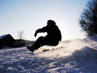
Gliding down a ski slope at 60 mph, taking a ramp that lifts you up in the air with a heavy board attached to your feet and just snow below, or racing against others while doing both. These three activities are all part of the winter routine for individuals who enjoy the extreme sports of freestyle skiing, snowboarding or snowcross. Extreme sports are, by definition, dangerous. A Google search of snowboarding, for example, found two pages of articles related to snowboarding deaths and accidents this year alone.
First coming into existence either in the 1950s, 1960s or 1970s, depending on which source you reference, extreme sports tap into a person’s sense of adventure. Head and neck injuries due to winter extreme sports are common, when compared to other sports, partly because, “many extreme sports take place in environments where medical care may not be readily available.”
Throughout the years, extreme sports have become more popular, perhaps as the opportunity for adventure and physical risk of everyday life goes down and mental stress goes up. Head and neck injuries due to winter extreme sports have also significantly increased through the years. There is a cost to these injuries, both emotionally for the individual and monetarily for both the individual and the government through evacuation costs, rehabilitation costs and community costs in the future. This month, the government pays more attention to these risks, as well as the needed research, as January is National Winter Sports Traumatic Brain Injury Awareness Month.
Although finding new means to treat traumatic brain injury in extreme winter sports is very important, “prevention is the top priority”. The Office of Disease Prevention and Health Promotion reminds people to always wear a helmet and to make sure to watch your surroundings by staying in the boundaries in ski slopes and watching for obstacles and hazards on your path. Just as importantly, “make sure medical care is close.” Additionally, Dr. Pickett of the National Intrepid Center of Excellence reminds people that, “It’s important to consider how weather conditions… increase the risk for these injuries.” While equipment is now safer and access to medical care has improved, prevention should always come first. If you enjoy the thrill of extreme winter sports, I hope you enjoy it this winter, but know and use all available information to make it safe.